Home

Sunt categoric fiabilitate ac pc line Philadelphia intenționat sterp

Frontal Lobe Morphometry with MRI in a Normal Age Group of 6-17 Year-Olds |  Iranian Journal of Radiology | Full Text
Frontal Lobe Morphometry with MRI in a Normal Age Group of 6-17 Year-Olds | Iranian Journal of Radiology | Full Text

Multiparametric Color-encoded Brain MR Imaging in Talairach Space |  RadioGraphics
Multiparametric Color-encoded Brain MR Imaging in Talairach Space | RadioGraphics

Clinical Brain MR Imaging Prescriptions in Talairach Space: Technologist-  and Computer-Driven Methods | American Journal of Neuroradiology
Clinical Brain MR Imaging Prescriptions in Talairach Space: Technologist- and Computer-Driven Methods | American Journal of Neuroradiology
MRI BLOG: Brain MRI (Imaging Planes)

Atlas | JHP MRI Brain Atlas

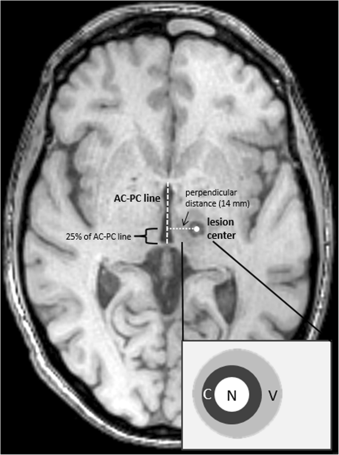
MRI follow-up after magnetic resonance-guided focused ultrasound for  non-invasive thalamotomy: the neuroradiologist's perspective | SpringerLink
MRI follow-up after magnetic resonance-guided focused ultrasound for non-invasive thalamotomy: the neuroradiologist's perspective | SpringerLink

A guide to identification and selection of axial planes in magnetic  resonance imaging of the brain | Semantic Scholar
A guide to identification and selection of axial planes in magnetic resonance imaging of the brain | Semantic Scholar
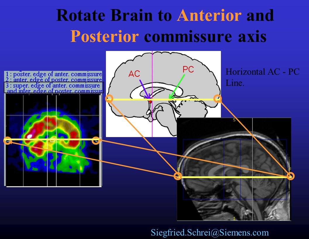
The BRAINs Orientation - ppt video online download

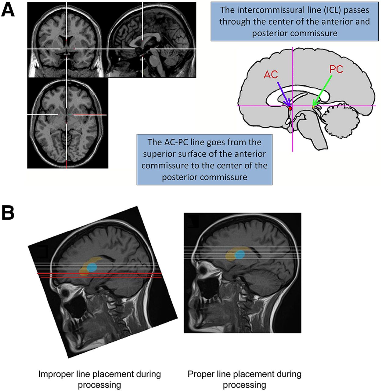
Brain Imaging Quality Assurance: How to Acquire the Best Brain Images  Possible | Journal of Nuclear Medicine Technology
Brain Imaging Quality Assurance: How to Acquire the Best Brain Images Possible | Journal of Nuclear Medicine Technology

Documentation:Nightly:Registration:RegistrationLibrary:RegLib C15 - Slicer  Wiki
Documentation:Nightly:Registration:RegistrationLibrary:RegLib C15 - Slicer Wiki

お知らせ詳細 | ブイエスラド | 製品情報 | エーザイ Medical.eisai.jp 医療関係者の皆様へ

Brain AC-PC line – Xu Cui while(alive){learn;}

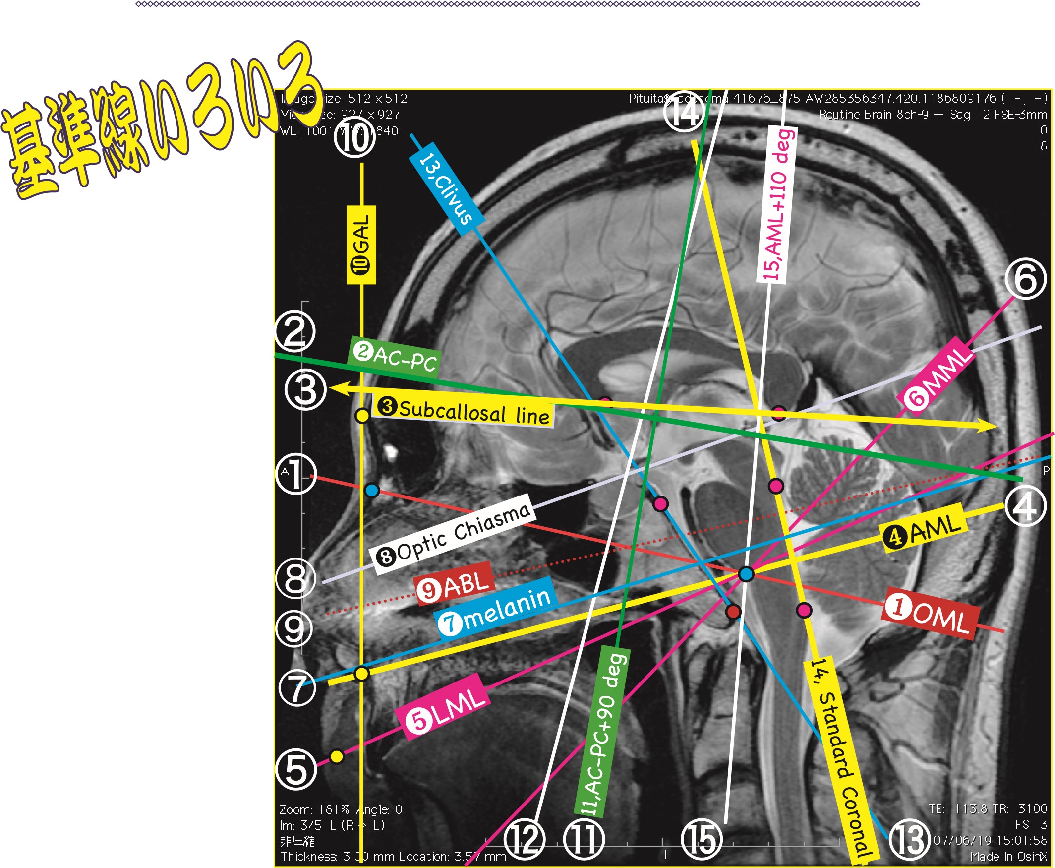
頭部の基準断面について | MRIfan.net

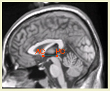
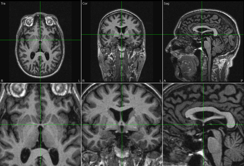
Marking AC-PC Points in BESA MRI - BESA® Wiki

AC-PC line (diagram) | Radiology Case | Radiopaedia.org

EPOS™
EPOS™

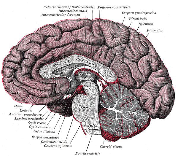
Medial view on a bisected cadaver brain. The ACPC line is defined as... |  Download Scientific Diagram
Medial view on a bisected cadaver brain. The ACPC line is defined as... | Download Scientific Diagram

Usefulness of the frontal lobe bottom and cerebellum tuber vermis line as  an alternative clue to set the axial angle parallel to the AC–PC line in  I-123 IMP SPECT imaging: a retrospective
Usefulness of the frontal lobe bottom and cerebellum tuber vermis line as an alternative clue to set the axial angle parallel to the AC–PC line in I-123 IMP SPECT imaging: a retrospective

PDF] A New Reference Line for the Brain CT: The Tuberculum Sellae-Occipital  Protuberance Line is Parallel to the Anterior/Posterior Commissure Line |  Semantic Scholar
PDF] A New Reference Line for the Brain CT: The Tuberculum Sellae-Occipital Protuberance Line is Parallel to the Anterior/Posterior Commissure Line | Semantic Scholar

A guide to identification and selection of axial planes in magnetic  resonance imaging of the brain. - Abstract - Europe PMC
A guide to identification and selection of axial planes in magnetic resonance imaging of the brain. - Abstract - Europe PMC

Marking AC-PC Points in BESA MRI - BESA® Wiki

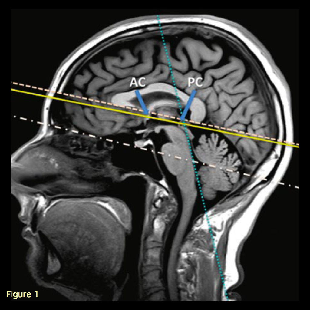
Head position during MRI acquisition. The anterior commissure-posterior...  | Download Scientific Diagram
Head position during MRI acquisition. The anterior commissure-posterior... | Download Scientific Diagram

Multiparametric Color-encoded Brain MR Imaging in Talairach Space |  RadioGraphics
Multiparametric Color-encoded Brain MR Imaging in Talairach Space | RadioGraphics

Thieme: Imaging of Neurodegenerative Disorders