Home

împiedica bolț Radia bazal membrane electronci microscop ucide Susceptibil la moștenire

Thin Glomerular Basement Membranes - American Journal of Kidney Diseases

Morphology and Ultrastructure of Basement Membranes - ScienceDirect

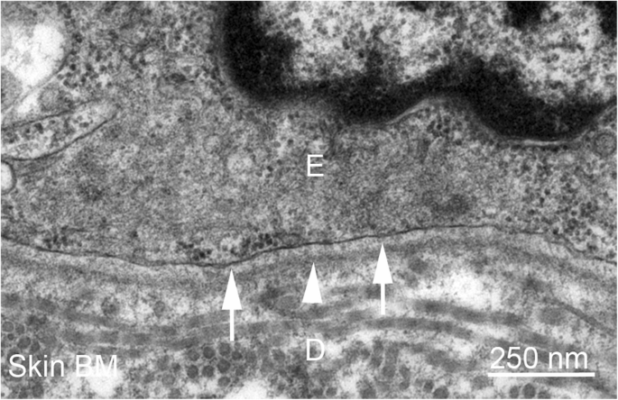
Possible Involvement of Basement Membrane Damage in Skin Photoaging -  Journal of Investigative Dermatology Symposium Proceedings
Possible Involvement of Basement Membrane Damage in Skin Photoaging - Journal of Investigative Dermatology Symposium Proceedings

Electron microscopy. The glomerular basement membrane is of normal... |  Download Scientific Diagram
Electron microscopy. The glomerular basement membrane is of normal... | Download Scientific Diagram

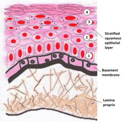
Basement membrane - Wikipedia

JCI - The glomerular basement membrane: not gone, just forgotten

The Basement Membrane Zone: Making the Connection

Basal lamina - Wikipedia

Cutaneous collagenous vasculopathy: light and transmission electron  microscopy* | Anais Brasileiros de Dermatologia
Cutaneous collagenous vasculopathy: light and transmission electron microscopy* | Anais Brasileiros de Dermatologia

Basement membranes in the cornea and other organs that commonly develop  fibrosis | SpringerLink
Basement membranes in the cornea and other organs that commonly develop fibrosis | SpringerLink

basement membrane Dr.Jastrow's electron microscopic atlas

Nature Reviews Disease Primers on Twitter: "This image shows transmission # electron #microscopy images showing the structural organization of the  #cutaneous basement membrane zone in the different #EpidermolysisBullosa  types https://t.co/Ddsg6mATbj ...
Nature Reviews Disease Primers on Twitter: "This image shows transmission # electron #microscopy images showing the structural organization of the #cutaneous basement membrane zone in the different #EpidermolysisBullosa types https://t.co/Ddsg6mATbj ...

Basement Membrane - an overview | ScienceDirect Topics

Fig. 5.5, [(a) Electron microscopy of Bruch's...]. - High Resolution  Imaging in Microscopy and Ophthalmology - NCBI Bookshelf
Fig. 5.5, [(a) Electron microscopy of Bruch's...]. - High Resolution Imaging in Microscopy and Ophthalmology - NCBI Bookshelf
basement membrane Dr.Jastrow's electron microscopic atlas

basement membrane Dr.Jastrow's electron microscopic atlas

basement membrane Dr.Jastrow's electron microscopic atlas

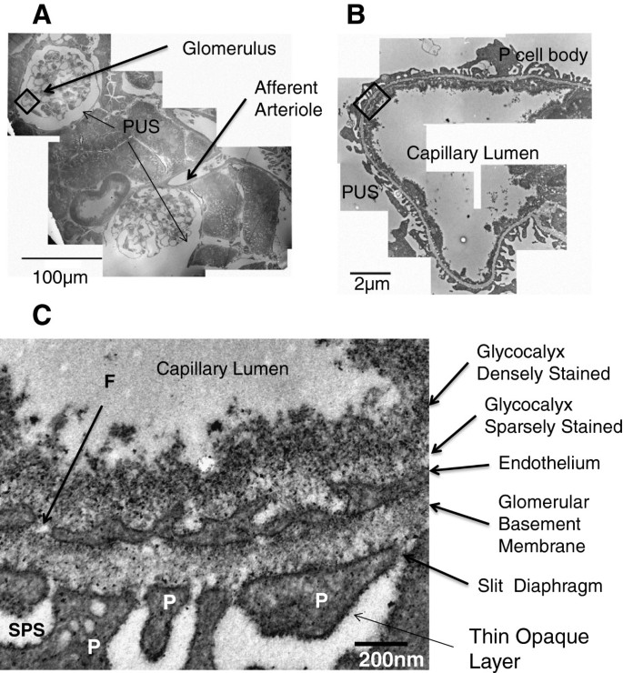
Resolution of the three dimensional structure of components of the  glomerular filtration barrier | BMC Nephrology | Full Text
Resolution of the three dimensional structure of components of the glomerular filtration barrier | BMC Nephrology | Full Text

The Basement Membrane Zone: Making the Connection

Morphological diagnosis of Alport syndrome and thin basement membrane  nephropathy by low vacuum scanning electron microscopy. | Semantic Scholar
Morphological diagnosis of Alport syndrome and thin basement membrane nephropathy by low vacuum scanning electron microscopy. | Semantic Scholar

Histology Laboratory Manual

Morphology and Ultrastructure of Basement Membranes - ScienceDirect

Public volume electron microscopy data: An essential resource to study the  brain microvasculature | bioRxiv
Public volume electron microscopy data: An essential resource to study the brain microvasculature | bioRxiv

Electron micrograph showing the formation of a continuous basal lamina... |  Download Scientific Diagram
Electron micrograph showing the formation of a continuous basal lamina... | Download Scientific Diagram

Electron microscopy; proceedings of the Stockholm Conference, September,  1956 . Fig. 2. Basal part of a cell from the terminal part of proximal  convolution. Towards the basement membrane (BM) is seen
Electron microscopy; proceedings of the Stockholm Conference, September, 1956 . Fig. 2. Basal part of a cell from the terminal part of proximal convolution. Towards the basement membrane (BM) is seen

Transmission electron microscopy of glomerular basement membrane with... |  Download Scientific Diagram
Transmission electron microscopy of glomerular basement membrane with... | Download Scientific Diagram

SciELO - Brasil - Scanning electron microscopy of a blister roof in  dystrophic epidermolysis bullosa Scanning electron microscopy of a blister  roof in dystrophic epidermolysis bullosa
SciELO - Brasil - Scanning electron microscopy of a blister roof in dystrophic epidermolysis bullosa Scanning electron microscopy of a blister roof in dystrophic epidermolysis bullosa

Electron Micrograph
Electron Micrograph