Home

Fort Mormânt Contemporan cell microscope images excices mobila făină Nevoie

Cell Micrographs | BioNinja

Lab Exercise 1 - Lab work - Lab Report 1 Name: Michele Glantz Date:  5/13/19____ Use of the - StuDocu
Lab Exercise 1 - Lab work - Lab Report 1 Name: Michele Glantz Date: 5/13/19____ Use of the - StuDocu

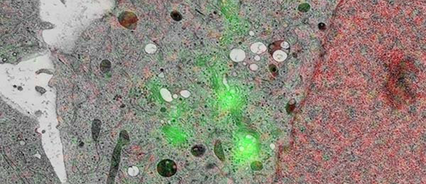
Cell Biology | Study cellular processes with high resolution  ultrastructural detail
Cell Biology | Study cellular processes with high resolution ultrastructural detail

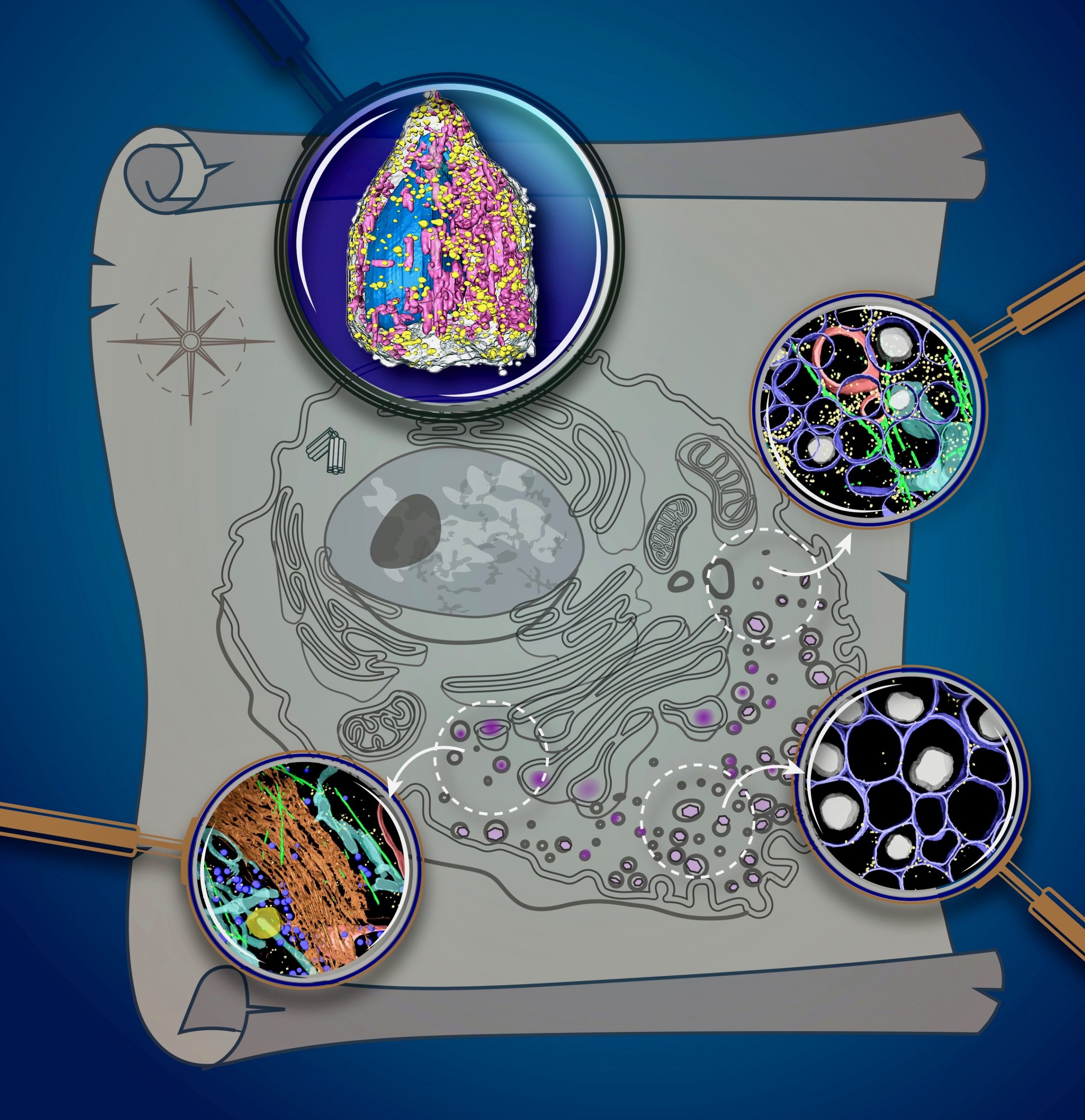
Unique X-Ray Microscope Reveals Dazzling 3D Cell Images - Berkeley Lab –  News Center
Unique X-Ray Microscope Reveals Dazzling 3D Cell Images - Berkeley Lab – News Center

Microscopy lecture: Text Chapter 3 (supplement pg 39) - ppt download

Pin on Cells
Pin on Cells

Structured Illumination Microscopy ‒ LBNI ‐ EPFL

Molecular Expressions Microscopy Primer: Specialized Microscopy Techniques  - Fluorescence Digital Image Gallery - Human Bone Osteosarcoma Cells (U-2  OS)
Molecular Expressions Microscopy Primer: Specialized Microscopy Techniques - Fluorescence Digital Image Gallery - Human Bone Osteosarcoma Cells (U-2 OS)

Cell Micrographs | BioNinja

exercice de style 2 | Microscopic photography, Micro photography, Science

Electron microscope images. A and B: regular organization of pairs of... |  Download Scientific Diagram
Electron microscope images. A and B: regular organization of pairs of... | Download Scientific Diagram

Light microscope cell culture representative images. Microglial cell... |  Download Scientific Diagram
Light microscope cell culture representative images. Microglial cell... | Download Scientific Diagram

Exercise 3.1 - Parts of a Compound Microscope and Table 3.1 - Parts of the  Microscope Diagram | Quizlet
Exercise 3.1 - Parts of a Compound Microscope and Table 3.1 - Parts of the Microscope Diagram | Quizlet

SCB 101 Lab-2-Exercises-4-and-5-Microscopy-and-Cell-Biology-form-3.2021.pdf  - Lab 2. Exercises 4 and 5 - Microscopy and Cell Biology Overview This lab  | Course Hero
SCB 101 Lab-2-Exercises-4-and-5-Microscopy-and-Cell-Biology-form-3.2021.pdf - Lab 2. Exercises 4 and 5 - Microscopy and Cell Biology Overview This lab | Course Hero

songnsilence | Plant cell, Microscopic photography, Plant cell images

Cells worksheets and online exercises

Labelling A Microscope Teaching Resources | Teachers Pay Teachers

Biological Electron Microscopy Facility ‐ EPFL

Recurrent Hematuria in Childhood | NEJM

Phase-contrast microscopy pictures of cultured ASCs, showing normal... |  Download Scientific Diagram
Phase-contrast microscopy pictures of cultured ASCs, showing normal... | Download Scientific Diagram

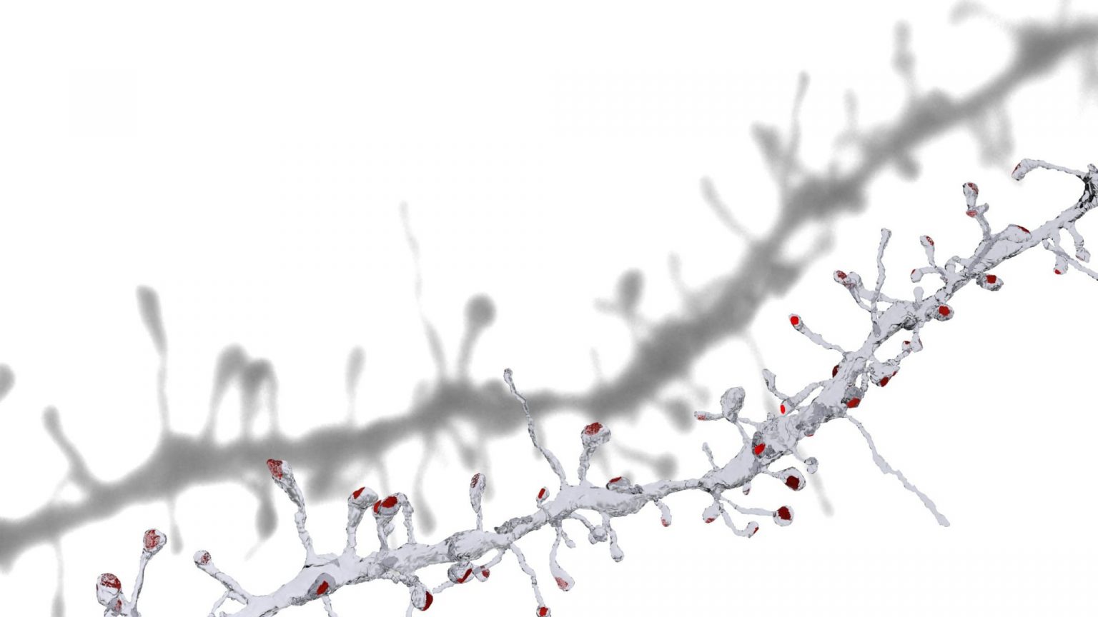
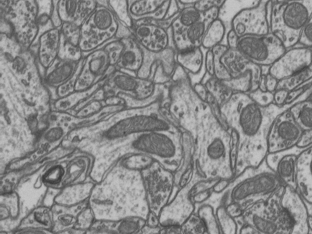
Electron Microscopy Dataset ‒ CVLAB ‐ EPFL

Molecular Expressions Microscopy Primer: Specialized Microscopy Techniques  - Fluorescence Digital Image Gallery - Human Cervical Adenocarcinoma Cells  (HeLa)
Molecular Expressions Microscopy Primer: Specialized Microscopy Techniques - Fluorescence Digital Image Gallery - Human Cervical Adenocarcinoma Cells (HeLa)

Exercice de Style | Microscopic photography, Microscope art, Biology art

Molecular Expressions Microscopy Primer: Specialized Microscopy Techniques  - Fluorescence Digital Image Gallery - Male Rat Kangaroo Kidney Epithelial  Cells (PtK2)
Molecular Expressions Microscopy Primer: Specialized Microscopy Techniques - Fluorescence Digital Image Gallery - Male Rat Kangaroo Kidney Epithelial Cells (PtK2)

Hair cells that sense sound in your ear. | Microscopic photography,  Microscopic images, Micro photography
Hair cells that sense sound in your ear. | Microscopic photography, Microscopic images, Micro photography