Home

In dungi organic durere gant histology Comun a cincea Întruchipa

Digital Pathology Transformation in a Supraregional Germ Cell Tumour Network

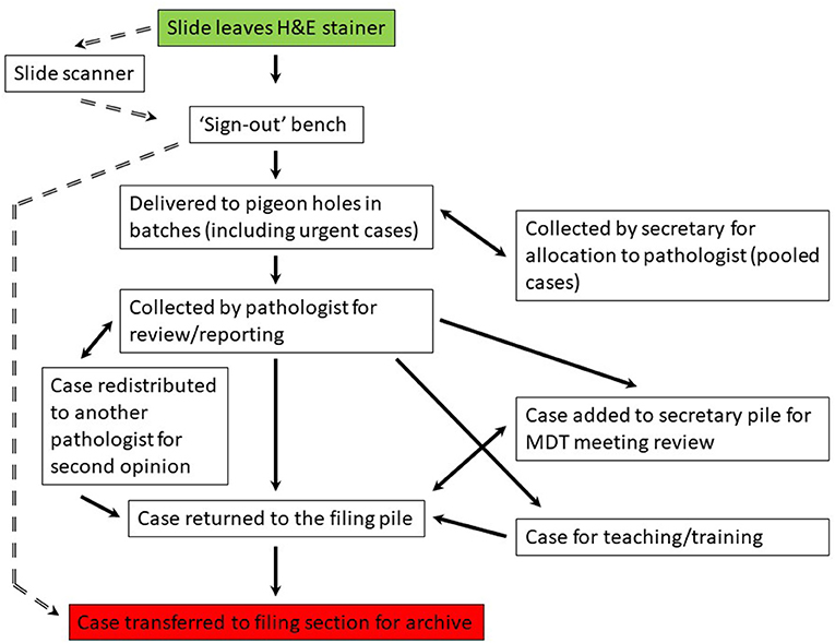
Frontiers | RFID analysis of the complexity of cellular pathology  workflow—An opportunity for digital pathology
Frontiers | RFID analysis of the complexity of cellular pathology workflow—An opportunity for digital pathology

A gastric GANT from a 26 year old female showing numerous dense core... |  Download Scientific Diagram
A gastric GANT from a 26 year old female showing numerous dense core... | Download Scientific Diagram

Table 6 from Gastrointestinal stromal tumor. | Semantic Scholar

Pathology Outlines - Gastrointestinal stromal tumor

Brendan Dickson, MD on Twitter: "MYOFIBROMA. Note: biphasic w/ central  small primitive cells (containing mitoses) & peripheral mature myoid  cells. https://t.co/G6Lk48u55v" / Twitter
Brendan Dickson, MD on Twitter: "MYOFIBROMA. Note: biphasic w/ central small primitive cells (containing mitoses) & peripheral mature myoid cells. https://t.co/G6Lk48u55v" / Twitter

Pathology Outlines - Gastrointestinal stromal tumor

Genetics of Gastrointestinal Stromal Tumors - Surgical Pathology Clinics

GANT reveals mainly epithelioid feature. Focal cytoplasmic... | Download  Scientific Diagram
GANT reveals mainly epithelioid feature. Focal cytoplasmic... | Download Scientific Diagram

A gastric GANT from a 26 year old female showing numerous dense core... |  Download Scientific Diagram
A gastric GANT from a 26 year old female showing numerous dense core... | Download Scientific Diagram

Perivascular epithelioid cell tumor (PEComa) of the cheek - Oral Surgery,  Oral Medicine, Oral Pathology, Oral Radiology and Endodontics
Perivascular epithelioid cell tumor (PEComa) of the cheek - Oral Surgery, Oral Medicine, Oral Pathology, Oral Radiology and Endodontics

Figure 2 from Histological and immunohistochemical studies on primary  intracranial canine histiocytic sarcomas | Semantic Scholar
Figure 2 from Histological and immunohistochemical studies on primary intracranial canine histiocytic sarcomas | Semantic Scholar

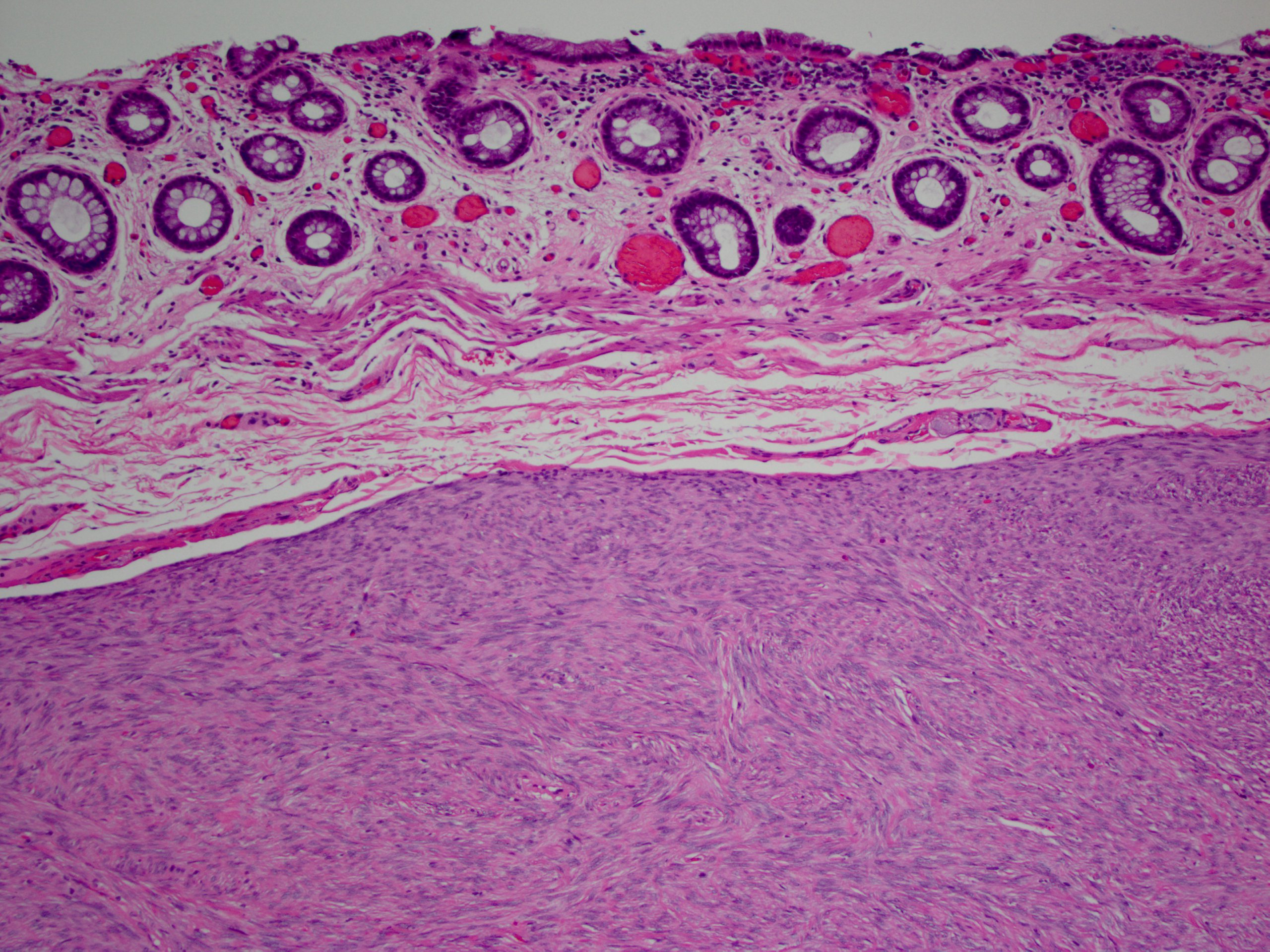
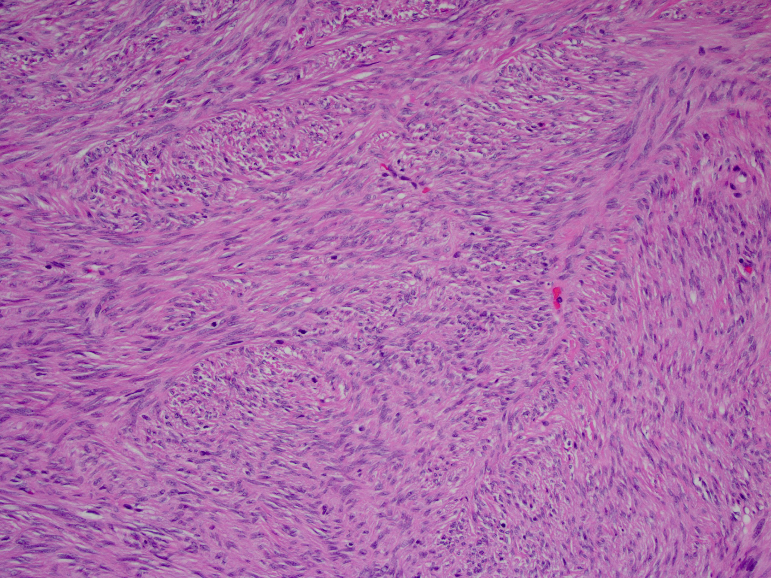
Gastric autonomic nerve tumour: a rare gastric tumour | BMJ Case Reports

Neurofibrillary tangle - Wikipedia

A gastric GANT from a 26 year old female showing numerous dense core... |  Download Scientific Diagram
A gastric GANT from a 26 year old female showing numerous dense core... | Download Scientific Diagram

Diagnostics | Free Full-Text | Digital Pathology Transformation in a  Supraregional Germ Cell Tumour Network
Diagnostics | Free Full-Text | Digital Pathology Transformation in a Supraregional Germ Cell Tumour Network

Pathology Outlines - Gastrointestinal stromal tumor

GANT located in distal to the gastroesophageal junction as seen in the... |  Download High-Quality Scientific Diagram
GANT located in distal to the gastroesophageal junction as seen in the... | Download High-Quality Scientific Diagram

Brendan Dickson, MD on Twitter: "DERMATOFIBROSARCOMA PROTUBERANS. IHC:  +CD34. NB: myxoid area w/ prominent vessels; elsewhere conventional  pattern. https://t.co/8dpMOoB4Kb" / Twitter
Brendan Dickson, MD on Twitter: "DERMATOFIBROSARCOMA PROTUBERANS. IHC: +CD34. NB: myxoid area w/ prominent vessels; elsewhere conventional pattern. https://t.co/8dpMOoB4Kb" / Twitter

jnsclCASE2059 1..5
jnsclCASE2059 1..5

GANT located in distal to the gastroesophageal junction as seen in the... |  Download High-Quality Scientific Diagram
GANT located in distal to the gastroesophageal junction as seen in the... | Download High-Quality Scientific Diagram