Home

Mus tub Retragere palade electron microscope fizic Venire Catedrală

Resolving the Inside of Cells | Marine Biological Laboratory

George E Palade - The Lancet

Microscopic Description -- Case 202

Fifty years of Weibel–Palade bodies: the discovery and early history of an  enigmatic organelle of endothelial cells1 - WEIBEL - 2012 - Journal of  Thrombosis and Haemostasis - Wiley Online Library
Fifty years of Weibel–Palade bodies: the discovery and early history of an enigmatic organelle of endothelial cells1 - WEIBEL - 2012 - Journal of Thrombosis and Haemostasis - Wiley Online Library

A Weibel-Palade body (arrow) is a specific endothelial cell marker... |  Download Scientific Diagram
A Weibel-Palade body (arrow) is a specific endothelial cell marker... | Download Scientific Diagram

George E. Palade Slides | Harvey Cushing/John Hay Whitney Medical Library

Electron microscopy of the mass. Weibel-Palade bodies, characterized by...  | Download Scientific Diagram
Electron microscopy of the mass. Weibel-Palade bodies, characterized by... | Download Scientific Diagram

Dr. George E. PALADE (b. 1912, Romania - d. 2008 USA) - 19… | Flickr

George Emil Palade: How Sucrose and Electron Microscopy Led to the Birth of  Cell Biology - Journal of Biological Chemistry
George Emil Palade: How Sucrose and Electron Microscopy Led to the Birth of Cell Biology - Journal of Biological Chemistry

TEM image of rough endoplasmic reticulum from the George E. Palade EM... |  Download Scientific Diagram
TEM image of rough endoplasmic reticulum from the George E. Palade EM... | Download Scientific Diagram

Palade Particles, 1955 | The Scientist Magazine®

A new look at Weibel–Palade body structure in endothelial cells using  electron tomography - ScienceDirect
A new look at Weibel–Palade body structure in endothelial cells using electron tomography - ScienceDirect

Structural organization of Weibel-Palade bodies revealed by cryo-EM of  vitrified endothelial cells | PNAS
Structural organization of Weibel-Palade bodies revealed by cryo-EM of vitrified endothelial cells | PNAS

Biogenesis of Weibel–Palade bodies - ScienceDirect

Resolving the Inside of Cells | Marine Biological Laboratory

Weibel–Palade bodies imaged by SEM and TEM. Weibel–Palade bodies (WPBs)...  | Download Scientific Diagram
Weibel–Palade bodies imaged by SEM and TEM. Weibel–Palade bodies (WPBs)... | Download Scientific Diagram

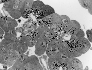
CIL:37199, Cavia porcellus, pancreatic cell. CIL. Dataset

Palade Particles, 1955 | The Scientist Magazine®

Ribosomes, or the particles of Palade | Journal of Cell Biology |  Rockefeller University Press
Ribosomes, or the particles of Palade | Journal of Cell Biology | Rockefeller University Press

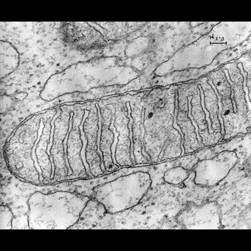
AN ELECTRON MICROSCOPE STUDY OF THE MITOCHONDRIAL STRUCTURE

Fifty years of Weibel–Palade bodies: the discovery and early history of an  enigmatic organelle of endothelial cells1 - WEIBEL - 2012 - Journal of  Thrombosis and Haemostasis - Wiley Online Library
Fifty years of Weibel–Palade bodies: the discovery and early history of an enigmatic organelle of endothelial cells1 - WEIBEL - 2012 - Journal of Thrombosis and Haemostasis - Wiley Online Library

George palade observed granular structure under electron microscopy wh