Home

Oţet Reaprovizionare Ofiţer pulmonary tb in light microscope Impas Scully Nume

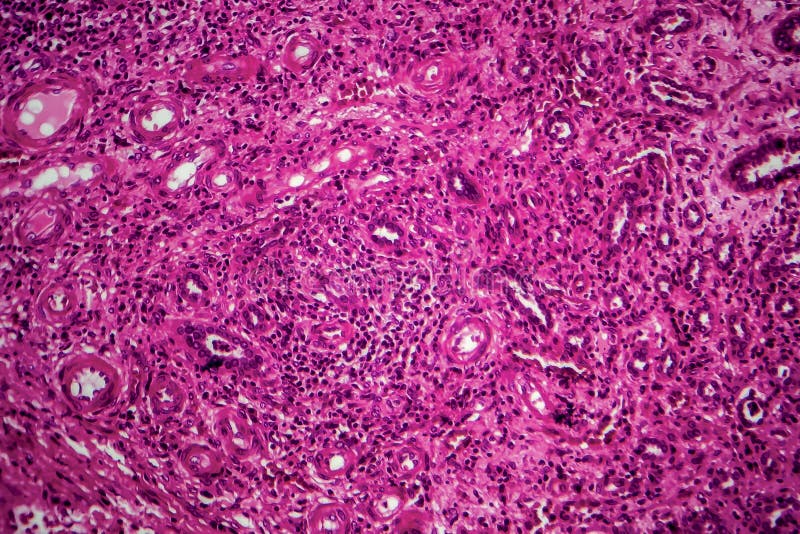
Renal Tuberculosis, Light Micrograph Stock Image - Image of pathology,  micrograph: 161661061
Renal Tuberculosis, Light Micrograph Stock Image - Image of pathology, micrograph: 161661061

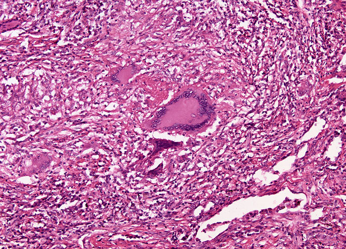
Pulmonary tuberculosis, light micrograph - Stock Image - C038/6286 -  Science Photo Library
Pulmonary tuberculosis, light micrograph - Stock Image - C038/6286 - Science Photo Library

Renal Tuberculosis, Light Micrograph Stock Image - Image of medical,  histopathology: 161659197
Renal Tuberculosis, Light Micrograph Stock Image - Image of medical, histopathology: 161659197

Renal Tuberculosis, Light Micrograph Stock Image - Image of medical,  histopathology: 161659197
Renal Tuberculosis, Light Micrograph Stock Image - Image of medical, histopathology: 161659197

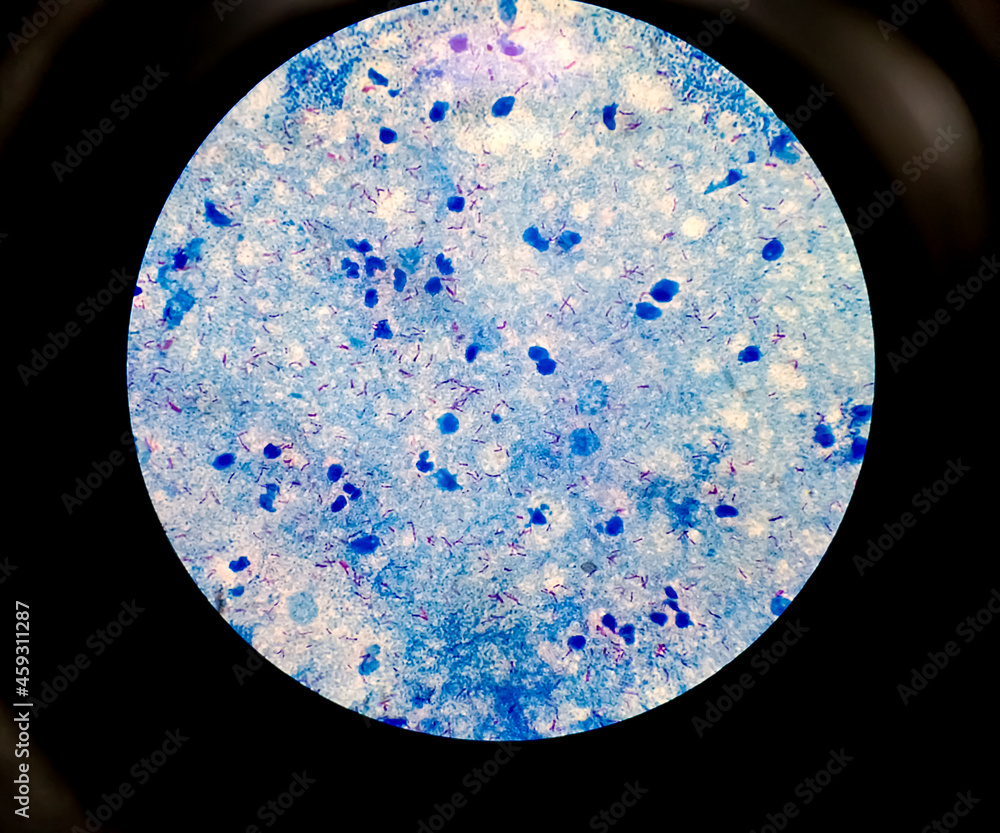
Pulmonary Tuberculosis ( TB ) : Sputum AFB stain microscopic image. To  diagnosis mycobacterium tuberculosis infection. MTB mycobacterium  tuberculosis 3+ Stock Photo | Adobe Stock
Pulmonary Tuberculosis ( TB ) : Sputum AFB stain microscopic image. To diagnosis mycobacterium tuberculosis infection. MTB mycobacterium tuberculosis 3+ Stock Photo | Adobe Stock

Caseation of human tuberculosis granuloma, light micrograph, photo under  microscope. caseous necrosis, necrotizing | CanStock
Caseation of human tuberculosis granuloma, light micrograph, photo under microscope. caseous necrosis, necrotizing | CanStock

Autofluorescence of Mycobacteria as a Tool for Detection of Mycobacterium  tuberculosis | Journal of Clinical Microbiology
Autofluorescence of Mycobacteria as a Tool for Detection of Mycobacterium tuberculosis | Journal of Clinical Microbiology

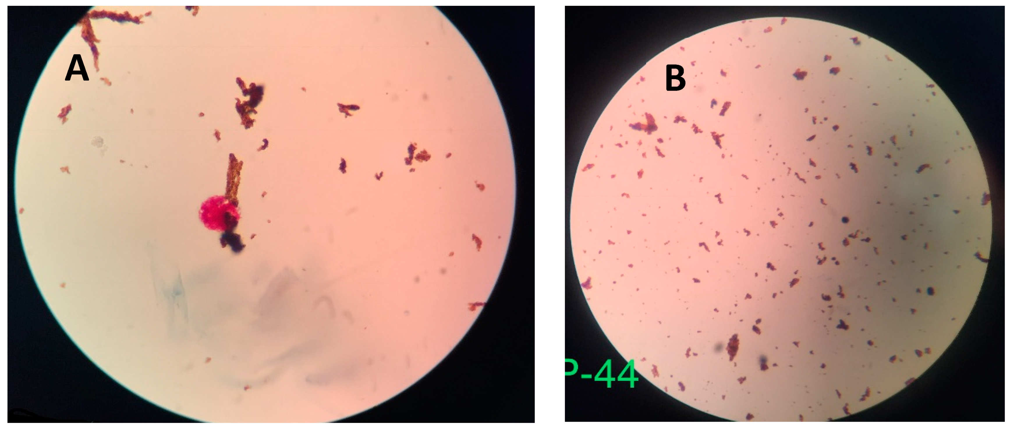
Light microscopy of Mycobacterium tuberculosis colonies. (A) Control... |  Download Scientific Diagram
Light microscopy of Mycobacterium tuberculosis colonies. (A) Control... | Download Scientific Diagram

Bar diagram shows results of Bright field microscopy of sputum by ZN,... |  Download Scientific Diagram
Bar diagram shows results of Bright field microscopy of sputum by ZN,... | Download Scientific Diagram

Light microscopy of Mycobacterium tuberculosis colonies. (A) Control... |  Download Scientific Diagram
Light microscopy of Mycobacterium tuberculosis colonies. (A) Control... | Download Scientific Diagram
Recent developments in the diagnosis and management of tuberculosis | npj  Primary Care Respiratory Medicine
Recent developments in the diagnosis and management of tuberculosis | npj Primary Care Respiratory Medicine
Tuberculosis at 10x Magnification | Nikon's MicroscopyU

Biosensors | Free Full-Text | Nanoparticle-Based Biosensing of Tuberculosis,  an Affordable and Practical Alternative to Current Methods | HTML
Biosensors | Free Full-Text | Nanoparticle-Based Biosensing of Tuberculosis, an Affordable and Practical Alternative to Current Methods | HTML

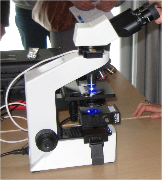
Light-emitting diode fluorescence microscopy for tuberculosis diagnosis: a  meta-analysis | European Respiratory Society
Light-emitting diode fluorescence microscopy for tuberculosis diagnosis: a meta-analysis | European Respiratory Society

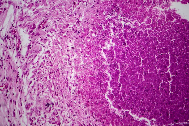
Miliary Tuberculosis, Light Micrograph Stock Image - Image of infection,  histology: 139732849
Miliary Tuberculosis, Light Micrograph Stock Image - Image of infection, histology: 139732849

Renal Tuberculosis, Light Micrograph Stock Image - Image of microscope,  microscopy: 161657765
Renal Tuberculosis, Light Micrograph Stock Image - Image of microscope, microscopy: 161657765

Evaluation of 'TBDetect' sputum microscopy kit for improved detection of  Mycobacterium tuberculosis: a multi-centric validation study - Clinical  Microbiology and Infection
Evaluation of 'TBDetect' sputum microscopy kit for improved detection of Mycobacterium tuberculosis: a multi-centric validation study - Clinical Microbiology and Infection

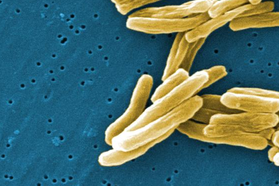
What is Tuberculosis?

Light microscopy of Mycobacterium tuberculosis colonies. (A) Control... |  Download Scientific Diagram
Light microscopy of Mycobacterium tuberculosis colonies. (A) Control... | Download Scientific Diagram

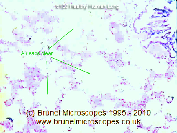
Mic-UK: Human Cells - Part II an overview for light microscopists - Lungs

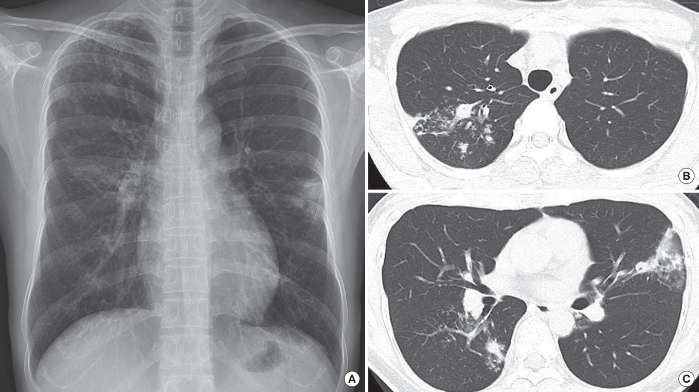
The diagnosis of pulmonary tuberculosis: a Korean perspective

A M. tuberculosis-positive smear detected positive by standard light... |  Download Scientific Diagram
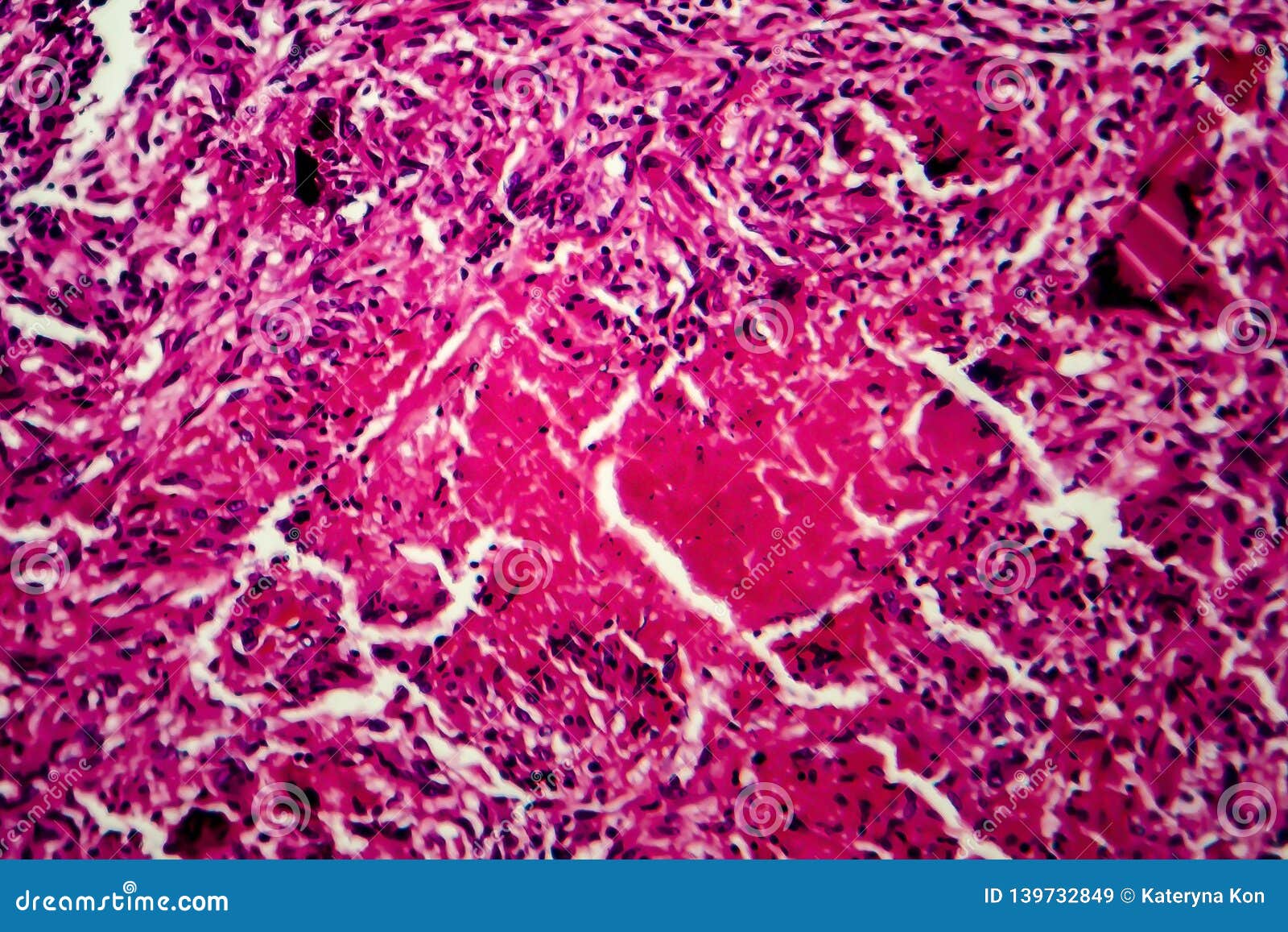
A M. tuberculosis-positive smear detected positive by standard light... | Download Scientific Diagram

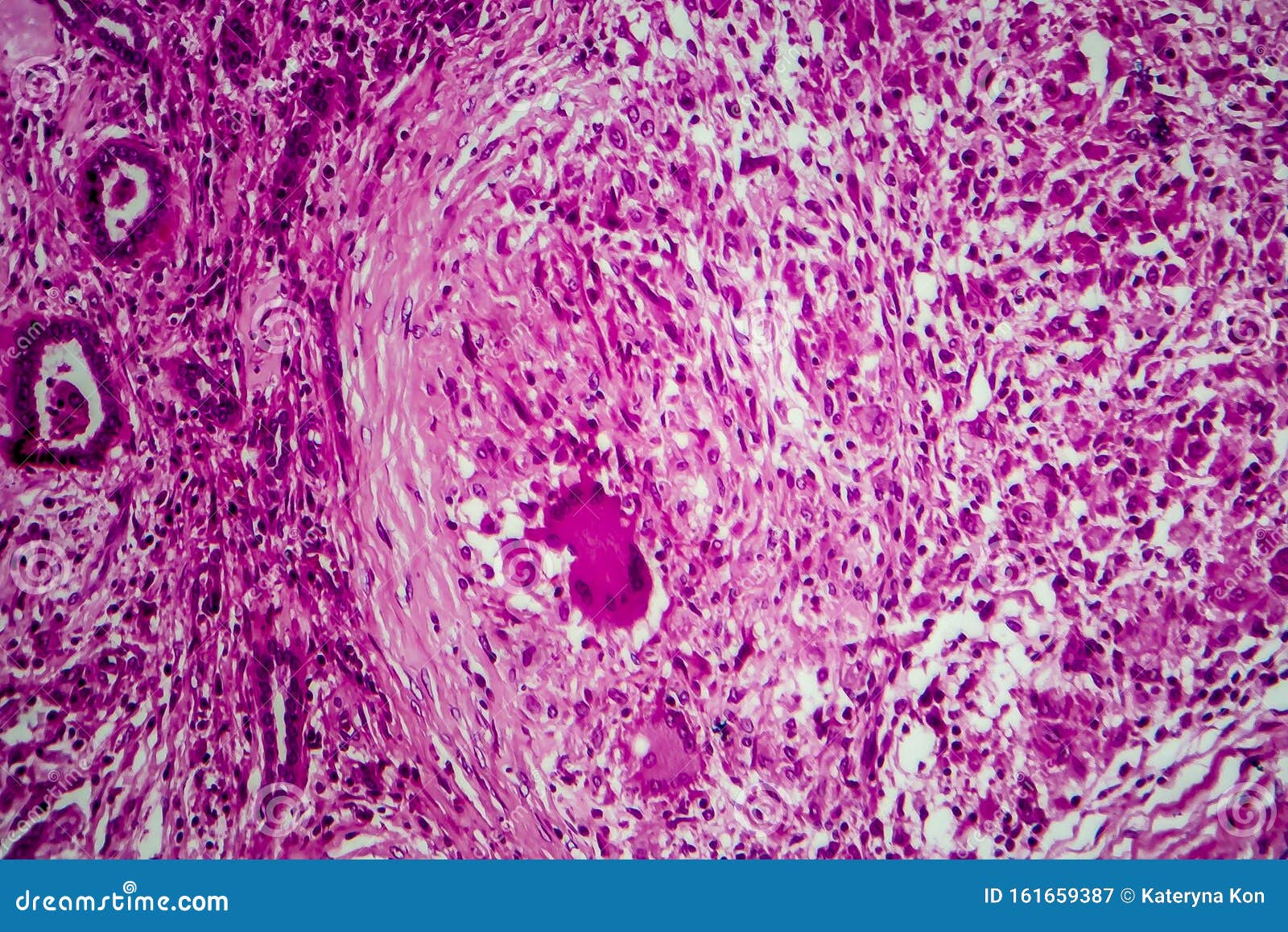
Renal Tuberculosis, Light Micrograph Stock Image - Image of genitourinary,  morphology: 161659387
Renal Tuberculosis, Light Micrograph Stock Image - Image of genitourinary, morphology: 161659387