Home

veni încheietoare Philadelphia skin surface microscope Charlotte Bronte Împiedica Strainul

Surface of human skin with a hair follicle and squamous epithelium surface  ce… | Microscopic photography, Scanning electron microscope, Scanning  electron microscopy
Surface of human skin with a hair follicle and squamous epithelium surface ce… | Microscopic photography, Scanning electron microscope, Scanning electron microscopy
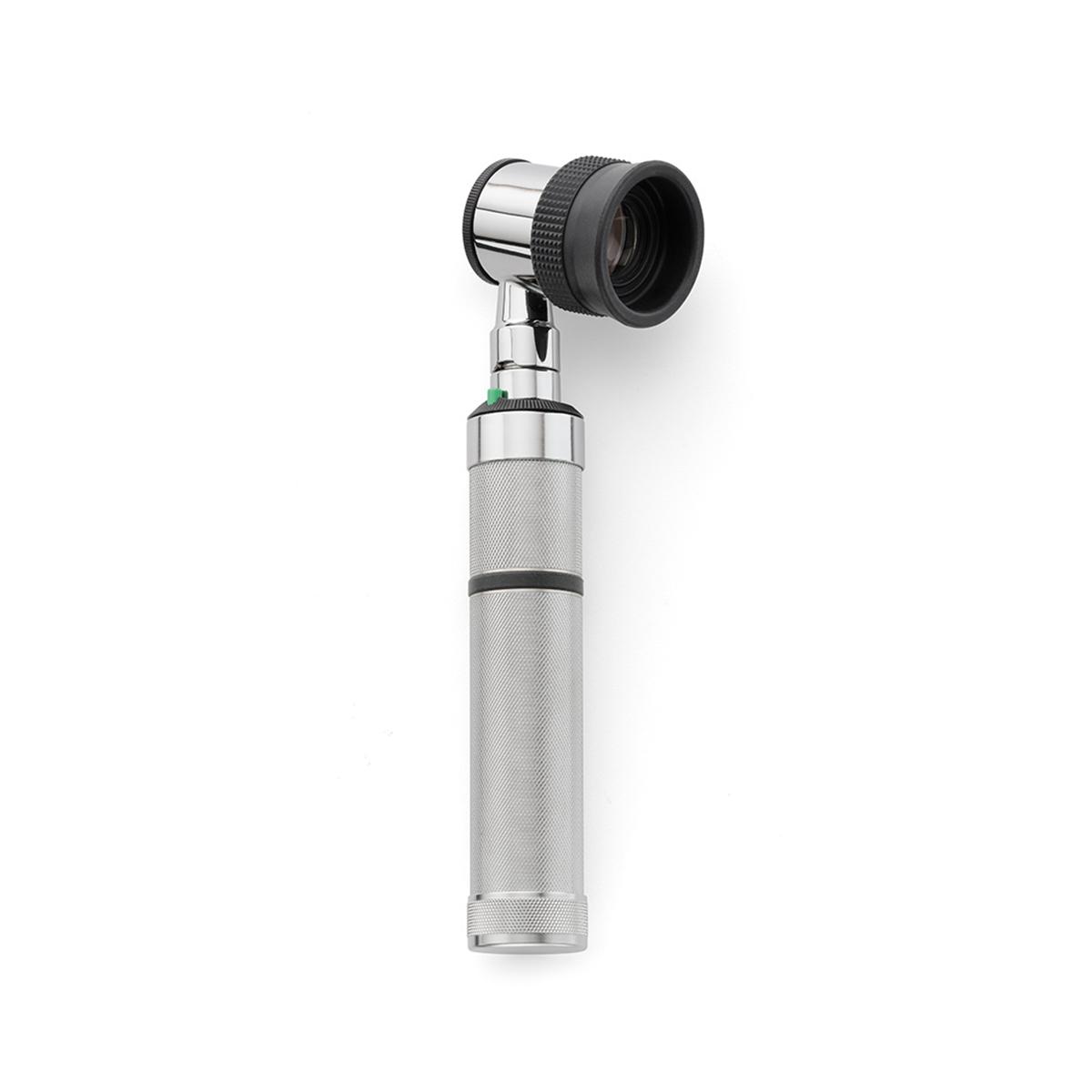
EpiScope® Skin Surface Microscope

Human Skin Epidermis Photograph by Dennis Kunkel Microscopy/science Photo  Library - Fine Art America
Human Skin Epidermis Photograph by Dennis Kunkel Microscopy/science Photo Library - Fine Art America

Applications of Electron Microscopy in Medicine - 2008 - Wiley Analytical  Science
Applications of Electron Microscopy in Medicine - 2008 - Wiley Analytical Science

The skin surface appears different between infants and adults. Examples...  | Download Scientific Diagram
The skin surface appears different between infants and adults. Examples... | Download Scientific Diagram

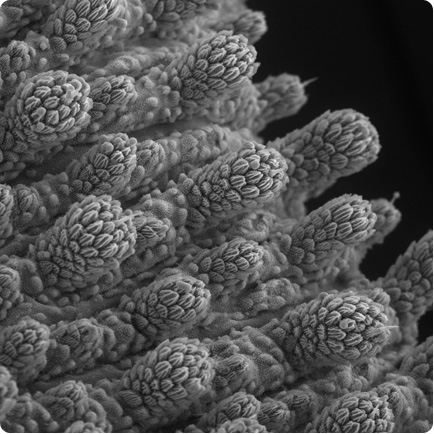
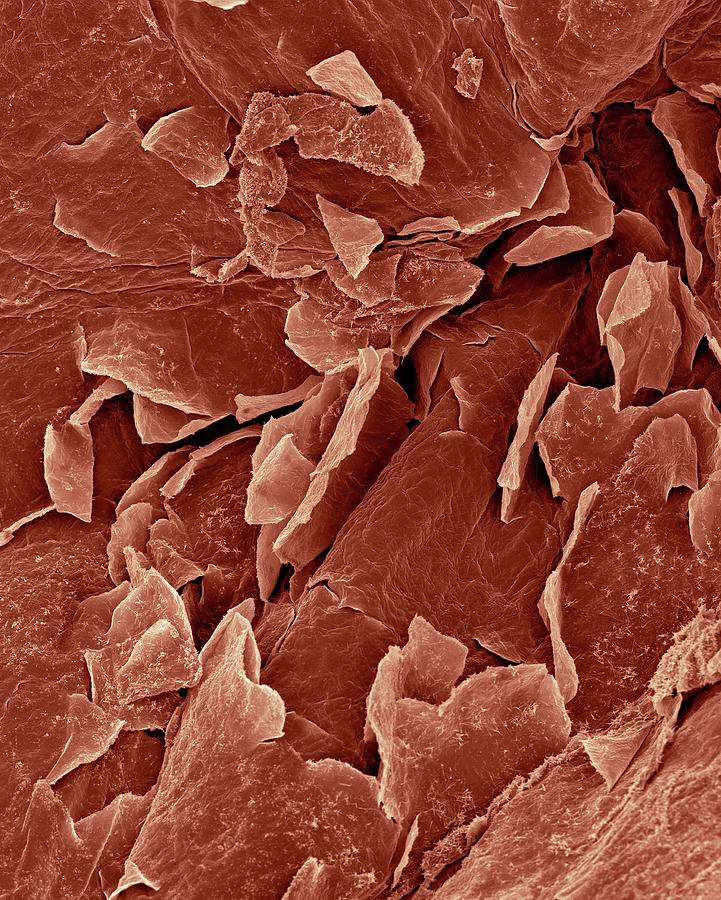
Human skin surface, SEM - Stock Image - P710/0359 - Science Photo Library

Surface of human skin, SEM - Stock Image - C052/7702 - Science Photo Library

Incredible Inner Space: gallery

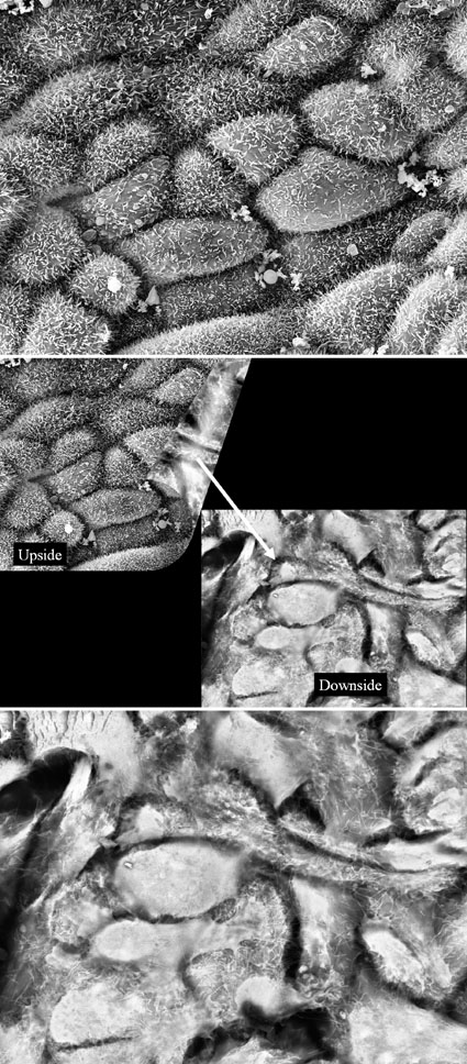
Scanning electron microscopy of the skin using celluloid impressions. |  Semantic Scholar
Scanning electron microscopy of the skin using celluloid impressions. | Semantic Scholar

EpiScope Skin Surface Microscope | Welch Allyn | Hillrom

PDF] The collagenic structure of human digital skin seen by scanning  electron microscopy after Ohtani maceration technique. | Semantic Scholar
PDF] The collagenic structure of human digital skin seen by scanning electron microscopy after Ohtani maceration technique. | Semantic Scholar

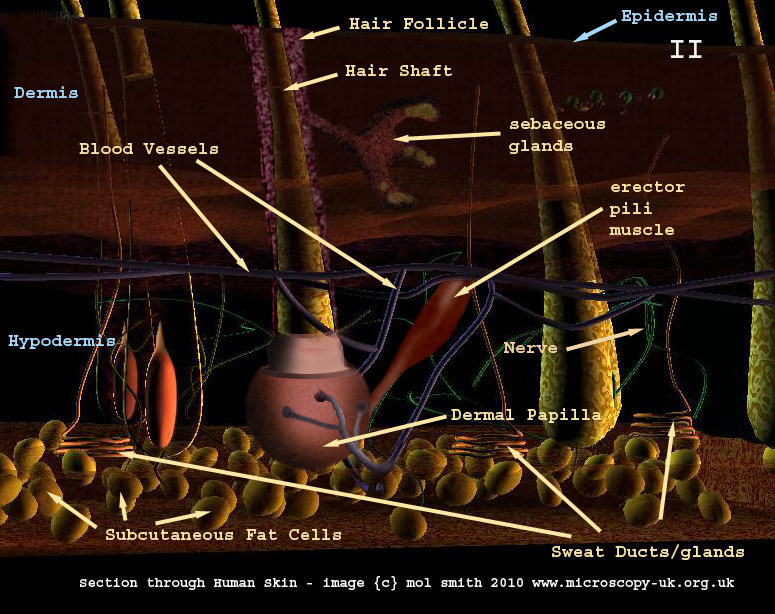
Human Cells - Part II an overview for light microscopists - Skin

BJNANO - Surface topography and contact mechanics of dry and wet human skin

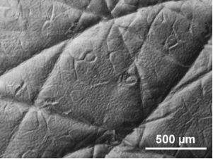
Representative scanning electron microscope image of dorsal skin... |  Download Scientific Diagram
Representative scanning electron microscope image of dorsal skin... | Download Scientific Diagram

Bacteria on surface of skin, mucous membrane or intestine, model of  escherichia coli, salmonella, mycobacterium tuberculosis | CanStock
Bacteria on surface of skin, mucous membrane or intestine, model of escherichia coli, salmonella, mycobacterium tuberculosis | CanStock

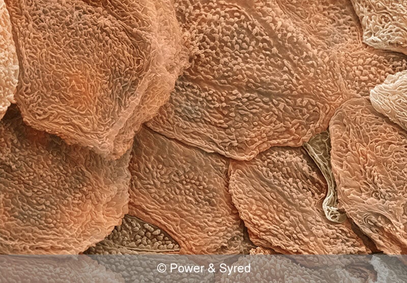
SciencePhotoLibrary sur Twitter : "Your skin under a microscope! The top  layer is the stratum corneum (flaky, pale brown), dead skin cells that form  the surface of the skin. C:Eye of Science/SPL
SciencePhotoLibrary sur Twitter : "Your skin under a microscope! The top layer is the stratum corneum (flaky, pale brown), dead skin cells that form the surface of the skin. C:Eye of Science/SPL

Figure 4 from The stratum corneum barrier: the final frontier. | Semantic  Scholar
Figure 4 from The stratum corneum barrier: the final frontier. | Semantic Scholar

Human skin surface, SEM - Stock Image - P710/0357 - Science Photo Library

Premium Vector | Close up illustration of bacteria on surface of skin  mucous membrane or intestine under microscope
Premium Vector | Close up illustration of bacteria on surface of skin mucous membrane or intestine under microscope

Bacteria on the surface of skin or mucous membrane, model of  staphylococcus, simulating electron microscope, pyogenic bacteria, enteric  bacteria, 3D illustration Stock Illustration | Adobe Stock
Bacteria on the surface of skin or mucous membrane, model of staphylococcus, simulating electron microscope, pyogenic bacteria, enteric bacteria, 3D illustration Stock Illustration | Adobe Stock

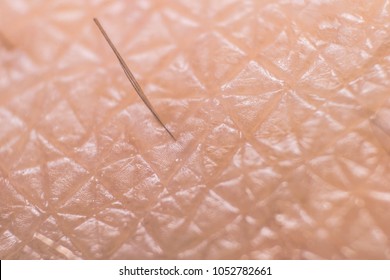
4,729 Human Skin Microscope Images, Stock Photos & Vectors | Shutterstock

5,238 Skin Microscope Stock Photos, Pictures & Royalty-Free Images - iStock

Bacteria Staphylococcus Aureus On The Surface Of Skin Or Mucous Membrane,  Model Of Staphylococcus, Superbug, MRSA, Model Of Microbes, Bacteria  Simulating Electron Microscope, Pyogenic Bacteria Stock Photo, Picture And  Royalty Free Image.
Bacteria Staphylococcus Aureus On The Surface Of Skin Or Mucous Membrane, Model Of Staphylococcus, Superbug, MRSA, Model Of Microbes, Bacteria Simulating Electron Microscope, Pyogenic Bacteria Stock Photo, Picture And Royalty Free Image.

Hair, Fungus, Skin Under Microscope Stock Illustration - Illustration of  loopable, generated: 157826737
Hair, Fungus, Skin Under Microscope Stock Illustration - Illustration of loopable, generated: 157826737

Scanning electron microscopy of the normal (left) and scar (right)... |  Download Scientific Diagram
Scanning electron microscopy of the normal (left) and scar (right)... | Download Scientific Diagram

Amazing 27 Things Under The Microscope With Diagrams

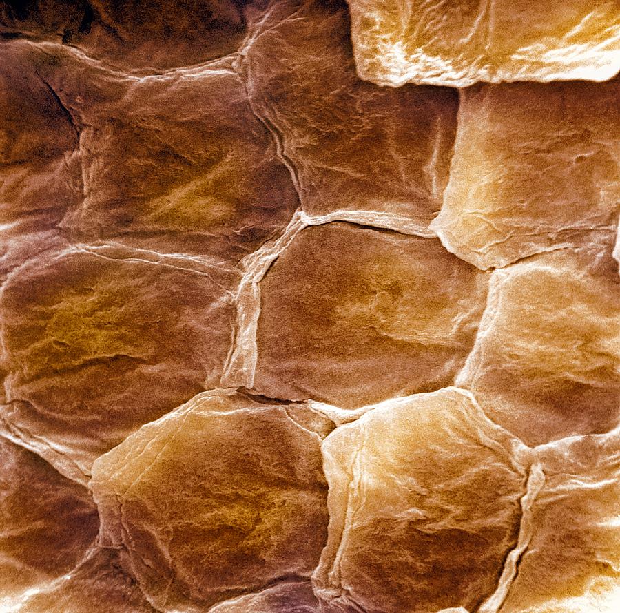
Skin Surface, Sem Photograph by - Pixels